How Does Physical Overtraining Affect the Brain?

Post by Shireen Parimoo

What is overtraining?

Every athlete, whether elite or recreational, strives to perform at their best. Behind every touchdown, race, or personal best is a dedicated training regimen. For example, marathon runners progressively increase their mileage week after week, with a mix of intense and easy training days. In the final “tapering” week before a race, they reduce the frequency and intensity (i.e. training load) of their running. The idea is to give the body enough time to recover from all the cumulative training in the weeks prior for optimal performance by race day.

Excessive exercise over a long period of time can lead to overtraining syndrome when there isn’t sufficient opportunity for recovery in between bouts of exercise. In addition to rest and recovery, adequate sleep, nutrition, and cross-training are also important for preventing overtraining. Functional overreaching is a common acute response to a high training load. At this stage, athletes tend to be fatigued from their training and feel like they need to exert more effort than usual to perform at their standard level. When the training load is reduced, the negative effects of functional overreaching on performance disappear within a week or so. In fact, athletes commonly experience a boost in their performance afterward, called “supercompensation”, which might explain the importance of tapering the week before a marathon.

What are the symptoms of overtraining?

Overtraining affects anywhere from 5-60% of professional athletes. If proper recovery is not built into a training regimen, then the effects of overreaching can persist and start to have a long-term impact not only on performance, but also on mood, lifestyle, and the brain. Some of the common symptoms include muscle soreness, fatigue, lowered immune response, depressive symptoms, cognitive problems like difficulty concentrating, and sleep issues.

There are three stages of overreaching and overtraining, with each stage becoming progressively worse and longer-lasting:

  1. non-functional overreaching: increased stress, fatigue, and muscle soreness along with poor sleep quality. Most of the time, this can be overcome by reducing training intensity and frequency and ensuring adequate sleep (1-3 weeks).

  2. sympathetic overtraining: further changes in fitness such as increased heart rate and muscular weakness, as well as higher cortisol levels and hormonal changes due to the prolonged stress (1-3 months). This is mostly observed in endurance athletes like long-distance runners.

  3. overtraining syndrome: a prolonged version of overtraining that can seriously alter the brain’s stress response and impact physical and mental health (6-12+ months).

How does regular exercise affect the brain?

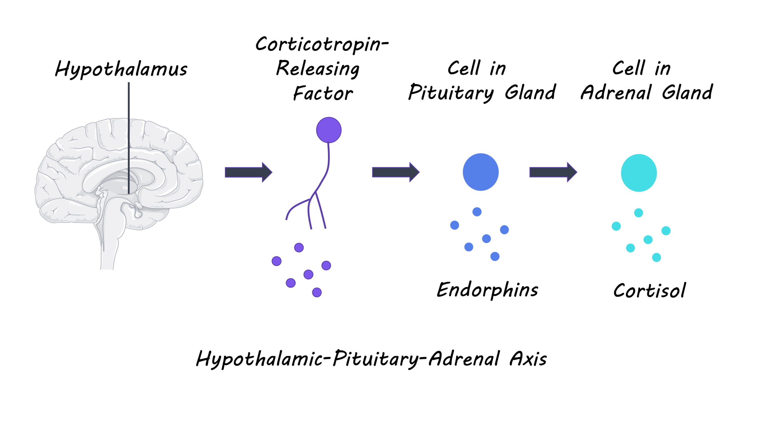
Neurotransmitter and hormonal imbalance contribute to overtraining. One way to think about overtraining is as a prolonged stress response to excessive exercise. Normally, the body’s stress response is regulated by the hypothalamic-pituitary-adrenal (HPA) axis. When a stressful event (i.e. an intense workout) occurs, noradrenaline levels increase and stimulate the hypothalamus in the brain. The hypothalamus releases the corticotropin-releasing factor, which stimulates the pituitary gland and also leads to increased heart rate. The pituitary gland then releases the adrenocorticotropic hormone, which stimulates the adrenal gland to produce the stress hormone cortisol. Cortisol suppresses the body’s immune response and triggers the “fight or flight” response by increasing blood glucose levels and heart rate. After a few hours, the cortisol circulating in the bloodstream inhibits the hypothalamus, terminating the stress response. Regular exercise helps the body adapt to physical stress and leads to decreased cortisol levels in response to future stressors. Importantly, this adaptation happens during the rest and recovery period. Exercise also increases serotonin levels in the brain and improves our sense of well-being. Serotonin is a neurotransmitter that affects mood, appetite, cognition, and sleep, among other important functions. Serotonin also regulates the stress response by acting on the HPA axis.

HPA_Feb23.png

How does overtraining affect the brain?

There is little opportunity for adaptation in between bouts of exercise without recovery periods, and excessive serotonin production might further contribute to HPA axis dysfunction. With overtraining, the body remains in a chronically activated stress response state that, in the long run, can lead to changes in the structure and function of various brain regions. Though research on the effects of overtraining on the brain and cognitive functions is somewhat scarce, there is ample evidence showing that a chronically activated stress response leads to changes in the brain. For example, prefrontal areas that help us with decision-making start to change in size and have different activation patterns in chronically stressed individuals. These individuals also start to rely on automatic rather than more effortful or strategic decision-making processes.

A recent study showed that excessive training affects decision-making processes. Endurance athletes completed an overreaching or normal training phase for three weeks. They were then brought into the lab to perform a reward-based decision-making task while undergoing an fMRI scan. The overreached athletes tended to choose smaller but immediate rewards over bigger rewards in the future, and they showed reduced activity in the prefrontal cortex when making decisions. Even though the authors could not – for ethical reasons – overtrain the athletes, this study provides an exciting avenue for future research on how overtraining affects brain functioning and cognition, which has several practical implications. Not only will it improve our understanding of how overtraining might impact the important day-to-day decisions of athletes, but this knowledge can be used to inform training plans that help ensure athletes get adequate rest and nutrition.

References

Armstrong & VanHeest. The unknown mechanism of the overtraining syndrome. Sports Medicine (2002). Access the original scientific publication here.

Cadegiani. Classical understanding of overtraining syndrome. In Overtraining Syndrome in Athletes (2020). Access the original scientific publication here.

Fulford & Harbuz. Chapter 1.3 – An introduction to the HPA axis. Techniques in the Behavioral and Neural Sciences (2005). Access the original scientific publication here.

Heisler et al. Serotonin activates the hypothalamic-pituitary-adrenal axis via serotonin 2C receptor stimulation. Journal of Neuroscience (2007). Access the original scientific publication here.

Kreher & Schwartz. Overtraining syndrome – A practical guide. Sports Health (2012). Access the original scientific publication here.

Lin & Kuo. Exercise benefits brain function: The monoamine connection. Brain Science (2013). Access the original scientific publication here.

Meeusen et al. Prevention, diagnosis and treatment of the overtraining syndrome: Joint consensus statement of the European College of Sport Science (ECSS) and the American College of Sports Medicine (ACSM). European Journal of Sport Science (2013). Access the original scientific publication here.

Portugal et al. Neuroscience of exercise: From neurobiology mechanisms to mental health. Neuropsychobiology (2013). Access the original scientific publication here

Soares et al. Stress-induced changes in human decision-making are reversible. Translational Psychiatry (2012). Access the original scientific publication here.

Wolff et al. Chronic stress, executive functioning, and real-life self-control: An experience sampling study. Journal of Personality (2020). Access the original scientific publication here.

How Sleep Helps Us Remember and Forget

Post by Amanda McFarlan

What’s the deal with sleep?

Humans spend approximately one third of their lives sleeping, so it is no surprise that we’re curious about it! Sleep has a wide variety of benefits, like repairing and regenerating tissues in the body, improving cognitive and physical performance, and consolidating memories. On the other hand, a chronic lack of sleep can put us at risk of developing health problems like cardiovascular disease, high blood pressure, diabetes, and depression. So, what happens when we sleep? Every night, when our heads hit the pillow, we enter into the first stage of ‘non-Rapid Eye Movement’ (non-REM) sleep. Non-REM sleep consists of 4 stages, with Stage 1 being the lightest sleep stage and Stage 4 being the deepest. Your body moves through the 4 stages of non-REM sleep and finally through REM sleep in a cycle that takes approximately 90 minutes, and this cycle is repeated throughout the night. Non-REM and REM sleep are characterized by different brain activity patterns, with non-REM sleep creating slow waves in its deepest stages, called ‘slow-wave sleep’, and REM sleep generating activity patterns that resemble wakefulness. The role of non-REM and REM sleep in the transfer and long-term storage of memories, known as memory consolidation, has been studied for many years. Here, we will discuss how sleep helps us remember or forget, as well as what goes wrong when we don’t sleep.

How does sleep help us remember?

Evidence strongly suggests that sleep is integral to memory consolidation. For example, a behavioural study, in which participants performed a visual task, a motor sequence task, and a motor adaptation task, found that participants’ performance was greatly improved if they had a full night’s sleep compared to those that did not sleep. The degree of performance improvement for each type of task was dependent on improved sleep in different stages in the sleep cycle. These findings suggest that non-REM and REM sleep both play an important role in memory consolidation. In line with this, other studies have shown that intensive learning of a new task is followed by increased time spent in REM sleep, resulting in subsequent task improvement, as well as the amplification of slow waves during non-REM sleep. Sleep results in a reactivation of cells in the hippocampus, which subsequently reactivate representations of memory in the cortex, also known as an engram. Over time, after many reactivations, these memories become distributed and consolidated within the cortex. 

sleep_jan26.png

Interestingly, research has shown that while we’re sleeping there is increased activity in the same hippocampal place cells (neurons that are activated when moving through specific locations in the environment) that were active throughout the day. This reactivation of hippocampal place cells during REM sleep follows a theta frequency band pattern of firing, hypothesized to be critical for memory consolidation. This hippocampal activity is mediated by neurons that release the neurotransmitter acetylcholine in the hippocampus. Acetylcholine, which plays a major role in altering the strength of synaptic connections, crucial for memory, is known to be elevated during REM sleep. REM sleep has also been associated with the upregulation of the expression of several calcium-dependent genes that are thought to be involved in synaptic plasticity and memory consolidation. 

Compared to REM sleep, the conditions in non-REM sleep are less ideal for promoting synaptic plasticity. For example, acetylcholine and calcium-dependent genes are expressed at low levels or are absent altogether during non-REM sleep. However, researchers have proposed that non-REM sleep might be important for the later stages of memory consolidation, rather than the initial conversion of short-term memories to long-term memories. In support of this, protein synthesis, which is required for long-term but not short-term potentiation (strengthening) of synapses, is increased during non-REM sleep. Therefore, the induction of protein synthesis during non-REM sleep may act to strengthen the synapses that were sufficiently potentiated during wakefulness. 

Although the majority of research on sleep and memory focuses on the role of the hippocampus in memory consolidation, a recent study has provided evidence that the thalamus might also play a role in memory consolidation during sleep. In this study, memory encoding (when memories are initially stored) during a visual task was shown to increase the activity of sensory relay nuclei of the thalamus in mice. Following a night of sleep, the primary visual cortex also showed evidence of a potentiated response to the visual task. Together, these findings suggest that task-related information may be passed from the thalamus to the primary visual cortex, resulting in the formation of a corresponding memory during sleep.

How does sleep help us forget?

Sleep research is centered around how we remember. However, sleep arguably plays just as important a role in the process of forgetting memories. The hippocampus serves as a temporary storage area for newly formed memories until they can be consolidated and integrated into long-term memory storage in the cortex. As a result, the hippocampus must be able to unlearn memories that have already been consolidated or memories that are not pertinent in order to store new memories. Research has shown that in addition to helping with memory consolidation, sleep is also important for unlearning memories. Studies in rats have shown that following sleep, there are widespread reductions in dendritic spines (protrusions on the dendrite that form synapses with nearby neurons) in the cortex as well as a reduction in receptors on glutamatergic neurons that are critical for memory and learning.

Norepinephrine and serotonin are two neurotransmitters in the brain that are associated with the enhancement of synaptic plasticity. During REM sleep, however, norepinephrine and serotonin signaling is suppressed, suggesting that REM sleep may allow for the depotentiation — or weakening — of synapses.  

What happens when we don’t sleep?

We all know how difficult it is to get through the day after a sleepless night. Suddenly, concentrating on what was previously a trivial task can become very challenging. Neuroimaging data has shown that sleep deprived individuals recruit more brain areas while performing the same cognitive task compared to individuals who slept normally. Moreover, brain imaging studies have revealed that hippocampal function is greatly reduced following one night of sleep deprivation, which suggests that losing sleep may actually disrupt our ability to learn new things. Sleep deprivation studies in rats have demonstrated the importance of REM sleep for learning as well as the induction and maintenance of long-term potentiation of synapses during learning. Additionally, REM sleep deprivation was shown to impair learning-dependent neurogenesis (the formation of new neurons) in the hippocampal dentate gyrus, which can impact future learning. The role of REM sleep for learning and memory is particularly relevant for individuals who are treated for depression with antidepressants, since these medications can greatly reduce the amount of time spent in REM sleep and may potentially have consequences on the efficacy of memory consolidation.

How can we get a good night’s sleep?

Given what we know about the role of sleep for learning and memory, it’s important to ensure that we get a good night’s sleep. However, with the challenges of daily life, this is not always an easy feat. First, it is important to establish a regular sleep schedule where you go to sleep and wake up around the same time each day, even when traveling or on the weekends. This habit can reinforce your body’s circadian rhythms, which helps your body to prepare for sleep and wakefulness more efficiently. Second, it is important to avoid using electronic devices before bed, like watching television or using your phone or tablet. The blue light that is emitted by these devices tricks our bodies into thinking it is daylight, and, as a result, our bodies produce lower levels of the hormone melatonin which promotes sleep. Third, use what you know about the science of sleep cycles to your advantage by timing your sleep in 90-minute intervals. For example, by setting your alarm for 7.5 hours of sleep (5 sleep cycles x 90 minutes each) you may actually feel more refreshed than if you slept for 8.5 hours and were awakened during the middle of a deep stage of sleep. Finally, avoiding caffeine and naps late in the afternoon or evening, as well as avoiding large meals or exercise right before bed may help to promote better sleep. 

Now, time to consolidate all of this learning with a good night’s sleep!

Feld, G.B., & Born, J. Sculpting memory during sleep: concurrent consolidation and forgetting. Current opinion in neurobiology, 44, 20–27 (2017). https://doi.org/10.1016/j.conb.2017.02.012

Klinzing, J.G., Niethard, N. & Born, J. Mechanisms of systems memory consolidation during sleep. Nat Neurosci 22, 1598–1610 (2019). https://doi.org/10.1038/s41593-019-0467-3

Poe, G. R., Walsh, C. M., & Bjorness, T. E. Cognitive neuroscience of sleep. Progress in brain research, 185, 1–19 (2010). https://doi.org/10.1016/B978-0-444-53702-7.00001-4

Stickgold, R. Sleep-dependent memory consolidation. Nature 437, 1272–1278 (2005). https://doi.org/10.1038/nature04286

How Should We Think About the Brain’s Response to Threat?

Post by Kasey Hemington

What's the science?

A threat is something with a high probability of causing either mental or physical damage. When we encounter a threat, many related processes occur in the brain, such as detecting the threat, learning to associate a cue with the impending threat, remembering what cues (and in what contexts) predict the threat, updating how or whether certain cues predict the threat over time, and deciding on the best behavioural response. These processes are typically studied independently and are each considered to be disrupted as distinct entities in threat-related disorders like anxiety and post-traumatic stress disorder (PTSD). This week in Trends in Cognitive Sciences, Levy and Schiller reviewed the neural basis of threat and proposed that we aim to understand threat in a more holistic manner; by studying the processes that make up the threat experience as interconnected phases of threat with common underlying neural computations.

What do we already know?

Though scientists often attempt to study them separately, it can be difficult to isolate the neural correlates of each aspect of the threat experience because each brain region known to be involved in these processes is involved in multiple processes. For example, the hippocampus, amygdala, and ventral striatum are involved not only in associative learning but also in decision-making, while areas of the prefrontal and parietal cortices are involved in decision-making but also learning. The insula is known to be involved in decision-making and learning, in addition to physiological reactivity, while the periaqueductal grey also plays a role in physiological reactivity, alongside providing threat-related signals to the amygdala. When it comes to understanding the brain’s response to threat, it may be more accurate to refer to the aforementioned regions as being part of one unified, global brain network.

What’s new?

Instead of studying each threat-related-process separately, the authors consider how different brain regions play a role in different ‘phases’ of the threat experience while asking the same neural computation-related question at each phase: in an uncertain and volatile environment, how does the brain use cues to predict outcomes?

For example, consider a person who witnesses a threatening event; the explosion of a blue car at close range. The authors divide this experience into five phases: 1) initial encounter (witnessing the explosion), 2) learning (that a blue car could signal danger), 3) post-association learning (e.g. learning whether the association should be generalized to cars of all colours), 4) memory retrieval and potential updating (remembering the danger in response to seeing another blue car, potentially stabilizing or destabilizing the threatening memory depending on the events and perception at the time of retrieval) and 5) decision making (e.g. choosing between whether to drive a car or ride a bicycle).  

kasey.jpg

The authors consider the neural correlates and clinical implications (for threat-related disorders) at each phase. 

Phase 1: As a threat becomes more imminent (for example, a predator moving into the field of view of its prey) there is a shift in brain activity from the prefrontal cortex to midbrain areas, and a corresponding shift in behavioural response from anxiety and fear to panic, freezing or fleeing. This pattern is mirrored in individuals with anxiety or PTSD; high anxiety is often experienced during anticipation of a threat before it is imminent. 

Phase 2: Learning the association between a cue and a threat occurs via prediction error in the brain; there is a difference between the expected and observed outcomes predicted by a cue, so the brain learns to update predictions. Synaptic plasticity in the amygdala results in the storage of threat memories. In individuals with anxiety disorders, learning may be overgeneralized in a maladaptive way to include cues that do not predict threat.

Phase 3: Extinction learning can counteract threat conditioning. It’s when repeated exposures elicit smaller and smaller responses to a stimulus over time. In the brain, the ventral tegmental area helps to compute a prediction error between expected and observed outcomes and sends a signal to other brain areas including the amygdala in order to update the memory with new extinction memories. In PTSD, defensive responses can linger following a threat for longer than they typically would.

Phase 4: When a memory is reactivated, this provides an opportunity to destabilize the memory (a cascade of cellular and molecular processes that put it in an unstable state) and potentially modify it in this unstable state. A clinical goal for PTSD and anxiety is to modify these threatening memories long-term, by reactivating the memory, alternating the memory to include a more adaptive emotional response, and ultimately altering the way an individual engages with the world. 

Phase 5: Decisions such as avoidance of a cue indicative of a threat are made based on subjective valuation of a potential outcome, for which the ventromedial prefrontal cortex and ventral striatum, in particular, are responsible. The uncertainty of the potential outcome is also encoded in the brain, including the ventral striatum, posterior parietal cortex, and anterior insula among other regions. Finally, the expected risk or reward and an individual’s overall tolerance for ambiguity are also weighed in the decision-making process. In PTSD and anxiety disorders, a decreased tolerance for ambiguity is observed.

What's the bottom line?

This review highlights learning, memory, and decision-making together as they relate to threat experiences and threat-related disorders. At the neural level, a response to threat can be thought of as computations that predict outcomes from cues. New associations become memories, which can be updated as behaviour and environments change. Finally, decisions can be made that incorporate these associations. This way of thinking about threats and related processes can help us to study the neural correlates of threat-related disorders like anxiety disorders and PTSD more holistically.

Levy and Schiller. Neural Computations of Threat. Trends in Cognitive Sciences (2020). Access the original scientific publication here.