Control of Ventral Visual System Neuronal Activity Using Deep Artificial Neural Networks

Post by Elisa Guma

What's the science?

The ventral visual stream is made up of six interconnected cortical brain areas responsible for transforming the light that strikes our retinas into visual representations, allowing us to recognize objects and their relationships in the world.  In order to better understand this complex visual processing system, neuroscientists have built computational models. Recent advances have allowed for better and more precise models using deep artificial neural networks (ANN) — which allow for each brain area within the ventral visual stream to be coded as a layer in the network. This week in Science, Bashivan and colleagues investigated the use and limitations of ANNs as models of neural processing in the nonhuman primate visual system V4 layer.

How did they do it?

In order to record neural activity in the visual cortex of awake macaques, the authors implanted micro-electrode arrays with 96 electrodes into the V4 layer of the left and right visual cortex of 3 monkeys. The area recorded by each of the 96 electrodes is referred to as a “neural site” and was only included in analyses if it maintained a stable response over the 3 days of experiments. On day 1, the authors determined the receptive field of each neural site using 640 naturalistic images and 370 “complex-curvature” images (computer generated) known to drive activity of V4 neurons. They were then able to use the neural response to 90% of these images to create a mapping from a “deep layer” (one of the processing layers between input and output in the neural network model) of the ANN to the neural responses; the remaining 10% of images were used to test accuracy of the model-to-brain mapping.

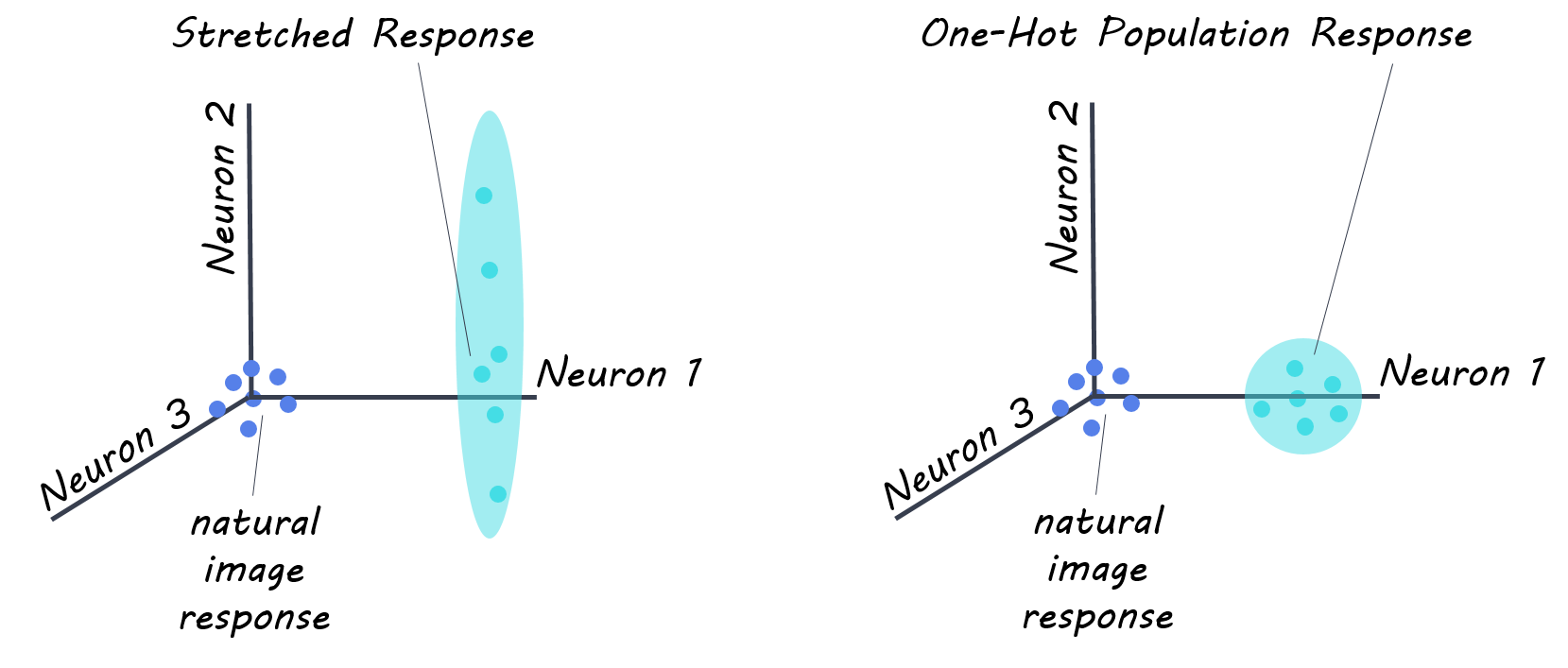
The next day, the authors performed a “stretch” control experiment, in which they instructed their model to drive the firing rate of one neural site as high as possible using a synthesized image (pattern of light) generated by the ANN. This control allowed for optimization of the response to each V4 site individually, without regard for the rest of the neural population. To investigate the system as a whole, on the third day, authors conducted a neural population state control referred to as “one-hot-population” control, to see if they ANN could generate images that would drive the response of one neural site, while simultaneously keeping the responses of all other sites low. These two controls allowed the authors to test the limitations of their model.

What did they find?

The authors recorded from 107 reliable sites for the ANN-mapping day, (52, 33, and 22 from each of the three monkeys respectively), 76 for the stretch control experiments (38, 19, 19 in each monkey), and 57 for the one-hot-population control experiment (38, 19 in each of two monkeys). First, they found their neural prediction model correctly predicted 89% of the V4 neural responses to the presented images. In their first control experiment, the “stretch” control, authors found that the algorithm generated images were able to produce firing rates 39% higher than the maximal firing rate occurring in neural sites when presented naturalistic images. This suggests that their ANN model was able to discover pixel arrangements that were better drivers of V4 visual cortex neurons. Finally, in the “one-hot-population” control experiment, the authors found that images did not achieve perfect population control (i.e. drive activity of one site only, while maintaining others at baseline), however, they did find that they were able to induce enhanced activity in the target site (by ~57% in 76% of sites) without too much of an increase in the off-target sites. This suggests that their ANN model is able to achieve better population control over the V4 neuronal population than previously possible.

Science.png

What's the impact?

These experiments show that ANN models can be used to generate images that drive firing rates at many V4 neural sites, and that these sites (even if they have overlapping receptive fields) can be partly independently controlled. The results suggest that the model has strong neuron-by-neuron functional similarity to the brain’s ventral visual stream (V4), suggesting that ANN models, although not perfect, may give new ability to find optimal stimuli to study neural systems in finer detail, unconstrained by limits of human language and intuition. This will in turn aid in our understanding of how the ventral visual system works.

DiCarlo_quote.jpg

Bashivan et al. Neural population control via deep image synthesis. Science (2019). Access the original scientific publication here.

Membrane Voltage Regulates Correlated Ion Channel Expression in Neurons

Post by Amanda McFarlan

What's the science?

The electrophysiological function of any given neuron is determined by the number and type of ion channels that are found in the neuron’s membrane. Neurons are able to maintain specific firing patterns by coordinating the expression of ion channels, although the underlying mechanisms of this process are still not well understood. Previous findings suggest that membrane voltage might play an important role in mediating firing patterns of neurons by providing homeostatic signals. This week in the Current Biology, Santin and Schulz investigated the role of membrane voltage in maintaining patterns of ion channel expression in the neuron.

How did they do it?

The authors began by assessing correlated mRNA expression patterns for 13 different ion channels. To do this, they dissected pyloric dilator neurons from the stomatogastric ganglion (a small motor circuit in decapod crustaceans that contains ~30 neurons each with distinct, identifiable characteristics) of adult male Jonah crabs. The mRNA expression of 13 different ion channels was quantified using single-cell PCR, followed by pairwise comparison in expression patterns between each of the 13 channels and every other channel. The first experiment had two conditions: control and silent. In the control condition, pyloric dilator neurons remained intact in their normal environment within the stomatogastric ganglion. In the silent condition, incubation in tetrodotoxin (sodium channel blocker) and the transection of the stomatogastric nerve (provides neuromodulatory inputs to the stomatostatic ganglion) resulted in pyloric dilator neurons that were deprived of all neural activity, synaptic input and neuromodulation.

In a second experiment, the authors investigated whether membrane voltage was important for maintaining correlated mRNA expression patterns of different ion channels. They measured mRNA expression levels in isolated pyloric dilator neurons for 8 hours under one of three conditions: control, silent or rescued. The control and silent conditions were the same as in the first experiment. In the rescued condition, pyloric dilator neurons were deprived from all synaptic input and neuromodulation as in the silent condition, however, the membrane potential was artificially restored to its original activity pattern by using a two-electrode voltage clamp. The authors used pairwise comparisons to assess mRNA expression pattern correlations across conditions.

What did they find?

The authors used pairwise comparisons to determine the relationship between mRNA expression levels in 13 ion channels. They showed that in the control group, 33 ion channel combinations (out of a possible 78 pairs of channels) had correlated patterns of mRNA expression. In the silent condition, they found a reduced correlation between mRNA expression levels among the 13 ion different ion channels, suggesting that neural activity, synaptic input and neuromodulation may play a critical role in regulating correlated expression of ion channel mRNA.

currentbiology.png

Next, the authors determined that 21 of the 33 pairs of ion channels with correlated mRNA expression patterns were shown to be significantly correlated in the control and rescued conditions, but not the silent condition, suggesting that the relationships between these ion channels are dependent on membrane voltage. The authors showed that in 4 out of 33 ion channel relationships, mRNA expression  correlations were present only in the control group. This finding suggests that these relationships were dependent on neuromodulatory feedback, rather than neural activity, since neuromodulatory inputs were not present in the silent or rescued conditions. Eight out of 33 ion channel interactions were unchanged across experimental groups, suggesting that they are not dependent on membrane voltage or neuromodulatory inputs. Finally, they found 5 new channel relationships that only appeared in the silent condition, suggesting that normal activity can not only influence relationships to form, but also suppress other interactions. Altogether, these findings suggest that membrane voltage may be an important factor in determining the correlation of ion channel expression.

What's the impact?

This is the first study to show that membrane voltage plays an important role in regulating mRNA expression patterns of ion channels, using a pyloric dilator neuron model. The authors also demonstrated that some ion channels have mRNA expression patterns that were not dependent on membrane voltage, which suggests that other mechanisms may be involved. Altogether, the direct link between ion channel activity and membrane voltage provides an important starting point for addressing other unanswered questions about ion channel activity patterns.  

Schulz_quote.jpg

Santin and Schulz. Membrane Voltage is a Direct Feedback Signal that Influences Correlated Ion Channel Expression in Neurons. Current Biology (2019). Access the original scientific publication here.

The Effect of Genetic Risk and Maternal Behavior on Children’s Amygdala Connectivity

Post by Shireen Parimoo

What's the science?

Depression and anxiety are internalizing disorders, which means that their symptoms are primarily experienced internally (e.g. sadness, loneliness) rather than being directed externally (e.g. impulsive behavior, bullying). Adverse childhood experiences like negative parental behavior are associated with a higher incidence of internalizing symptoms and depression later in life. Depression has also been linked to disrupted functioning of the amygdala and the hypothalamic-pituitary-adrenal (HPA) axis, a set of brain regions that control the body’s reaction to stress. Together, negative childhood experiences and having certain genes associated with the HPA axis are related to having depression, but the mechanism of this interaction is unclear. This week in NeuroImage, Pozzi and colleagues used functional magnetic resonance imaging (fMRI) to delineate the relationship between HPA genetic risk, parental behavior, amygdala activation, and children’s depressive symptoms.

How did they do it?

Eighty children aged 8 - 9 years old participated in a longitudinal study with two time points. At the first time point, the authors recorded interactions between the children and their mother and 18 months later, the children performed an emotional processing task while undergoing fMRI scanning. The mother-child interactions were each 15 minutes long and consisted of an event-planning interaction and a problem-solving interaction. The mothers’ behavior was categorized into negative (e.g. anger) and positive (e.g. listening) behavior during each interaction. Saliva samples were also collected at the first time point to assess genetic risk, yielding a composite HPA genetic risk score based on the number of HPA-related genes that they possessed.

At the second time point, children completed questionnaires assessing internalizing symptoms, depression, and anxiety, while their mothers completed questionnaires assessing maternal depression and their children’s internalizing symptoms. The children also completed an emotional face-matching task in which they had to match the gender of an angry or fearful target face with one of two other faces. In the control condition, participants matched shapes instead of faces. The authors first examined task-related activity in the amygdala when participants performed the face compared to the shape matching task. They then performed a generalized psychophysiological interaction (gPPI) analysis to determine how the connectivity between the amygdala and other regions of the brain were related to the interaction between genetic risk, child functioning, and maternal behavior.

What did they find?

There were no direct associations between amygdala activation during the emotional face-matching task, and the interaction between genetic risk, and maternal behavior. Genetic risk moderated the relationship between negative maternal behaviors and brain connectivity. Specifically, higher genetic risk was linked to greater amygdala connectivity with the right superior frontal gyrus when mothers exhibited more negative behaviors during the problem-solving task. Conversely, if mothers exhibited more negative behaviors but the child’s genetic risk was low, the connectivity between the amygdala and the right superior frontal gyrus was also lower. During the event planning task, negative maternal behavior was associated with greater connectivity between the amygdala and the postcentral gyrus in the presence of high genetic risk, but reduced amygdala connectivity with fronto-parietal regions when genetic risk was low. This suggests that how mothers interact with their children affects the children’s amygdala’s connectivity in different ways depending on the level of genetic risk for HPA axis dysregulation.

Pozzi_image.jpg

Finally, genetic risk was not directly associated with children’s internalizing or depressive symptoms. However, there was an indirect relationship between genetic risk and child functioning that was mediated by the amygdala’s connectivity to the precuneus. Higher genetic risk was linked to more internalizing symptoms in children via higher amygdala-precuneus connectivity.

What's the impact?

This study is the first to show that genetic risk influences the effect that negative maternal behavior has on brain activity (connectivity) related to children’s emotion processing. The results further illustrate that altered brain functioning underlies the interaction between genetic risk factors and depressive symptoms. These findings provide deeper insight into how genetic and environmental variables might contribute to the development of internalizing disorders such as depression and anxiety.

Pozzi_quote.jpg

Pozzi et al. Interaction between hypothalamic-pituitary-adrenal axis genetic variation and maternal behavior in the prediction of amygdala connectivity in children. NeuroImage (2019). Access the original scientific publication here.