Mediated Learning is Dependent on Type 1 Cannabinoid Receptors

Post by Sarah Hill

What's the science?

CB1 receptors bind cannabinoids (a class of chemical compounds) produced within the body (endocannabinoids) and are known to be involved in learning and memory. Binding of CB1R to cannabinoids originating outside of the body, for example during consumption of marijuana, can cause cognitive impairment. In contrast, endocannabinoid binding to CB1R within the hippocampus is involved in direct associative learning (a simple form of learning where an association is made between a certain stimulus and an outcome, such as the pairing of a sound with a subsequent food reward). Whether CB1R and the endocannabinoid system in general are involved in other higher-order forms of learning, such as ‘mediated learning’, is not known. Mediated learning is when a stimulus is paired with an outcome indirectly through association with another stimulus (also known as an incidental association). This week in Neuron, Busquets-Garcia and colleagues demonstrate that hippocampal CB1Rs are specifically required for mediated learning.

How did they do it?

The authors carried out a sensory preconditioning task in mice with a number of transgenic and pharmacological interventions. The sensory preconditioning procedure consisted of three phases: 1) Two low-salience stimuli (such as odor and taste) were presented to male mice simultaneously to promote formation of an incidental association between the two (pre-conditioning phase) 2) One of the original stimuli was directly paired with either an aversive or a reward reinforcer (conditioning phase), increasing the salience of the outcome-associated stimulus and indirectly pairing the other stimulus with the same outcome 3) Mice were presented with either the directly-paired or the indirectly-paired stimuli in order to evaluate direct associative learning or mediated learning respectively (test phase). In the first variation of sensory preconditioning, the authors paired odor and taste with an aversive outcome (gastric malaise). To ensure that their results were not restricted to odor and taste stimuli, nor to an aversive outcome, the authors carried out an additional sensory preconditioning task by pairing two new stimuli, a light and a sound, with a food pellet reward outcome.  

sarah1800.png

As a basic test of CB1R activity in mediated learning, sensory preconditioning was first carried out using mice who had CB1R deleted or 'knocked-out' on a brain-wide level. Next, to determine the timeframe of CB1R activity during mediated learning, the authors administered a CB1R antagonist (i.e. blocker) at different time points throughout the task, either before the preconditioning phase or the test phase. To assess which brain region CB1R-dependent mediated learning occurs within, they repeated the experimental procedure after deleting CB1R within the hippocampus. Finally, to identify the neurons involved in CB1R-dependent mediated learning, the authors carried out the sensory preconditioning task in mice lacking CB1R specifically in forebrain GABAergic inhibitory interneurons. Because CB1R helps to suppress GABAergic inhibitory neurotransmission, the authors hypothesized that CB1R inhibition of GABA neuron firing may be critical for incidental associations and mediated learning to occur. To test this, they infused a viral vector expressing inhibitory designer receptors exclusively activated by designer drugs (DREADD) to either inhibit or excite GABAergic neurons in the hippocampi of mice lacking CBR1 before preconditioning. This allowed them to observe the impact of activating or inhibiting GABAergic neurons on mediated learning.

What did they find?

Wild-type (control) mice that underwent sensory preconditioning consumed reduced amounts of tastes or odors that were indirectly or directly paired with gastric malaise, suggesting that mediated and associative (direct) aversion learning had occurred. Likewise, increased reward-seeking behavior was observed in response to stimuli when two associated stimuli were directly- and indirectly-paired with a reward outcome. However, the results did not hold for indirect-pairing of a stimulus-outcome duo when CB1R knock-out mice or CB1R antagonist-dosed mice underwent sensory preconditioning (direct-pairing was unaffected). This suggests that CB1 receptors are required for higher-order/mediated learning to occur. Administration of a CBR1 antagonist (blocker) at different timepoints revealed that CB1Rs are specifically activated during the formation of incidental associations, but not during expression of mediated aversive or reward-seeking behaviors. Additional transgenic and viral approaches further confirmed that CB1 receptors involved in this mode of learning are uniquely expressed by GABAergic interneurons within the hippocampus.

Suppression of hippocampal GABAergic signaling (accomplished using inhibitory DREADDs) in mice lacking CBR1 in the hippocampus, was sufficient to recover mediated learning deficits (normally observed in animals lacking CB1R inhibitory function). Conversely, excitatory DREADDs used to activate inhibitory GABAergic transmission, effectively overriding suppression by CB1R, resulted in reduced consumption of the indirectly-paired taste stimulus but not the directly-paired stimulus. Taken together, these findings indicate that excess inhibitory signaling during preconditioning disrupts the formation of incidental associations, blocking mediated learning. Therefore, CB1R are necessary to regulate the activity of GABAergic interneurons. The authors performed a follow-up experiment to show that the GABAergic neurons involved are not parvalbumin positive interneurons (approximately half of GABAergic neurons in the hippocampus). This suggests that the remaining half, presumably interneurons containing cholecystokinin (CCK), are the neuronal subpopulation responsible for driving mediated learning.       

What's the impact?

This study identified a unique role for Type 1 cannabinoid receptors in a form of higher-order learning called mediated learning. Specifically, CB1R expressed by non-parvalbumin GABAergic interneurons within the hippocampus contribute to this learning process and are activated during formation of incidental associations. As CB1R has been implicated in a number of psychiatric and neurological disorders, this is an important finding to consider when designing CB1R-targeting therapeutic strategies.

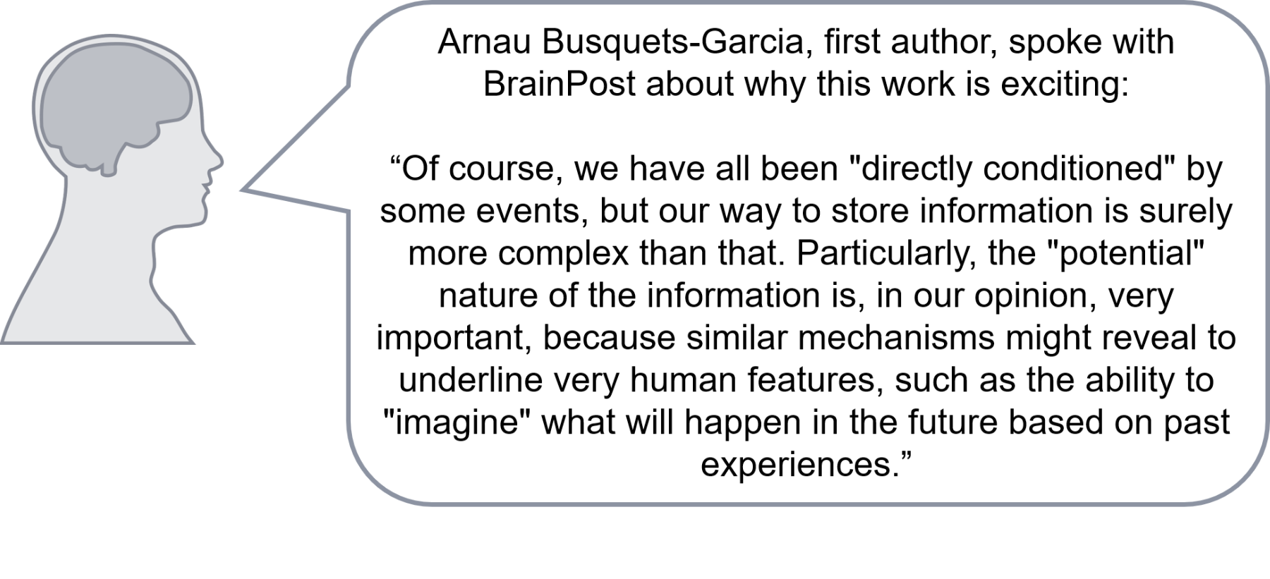
sarahquote1800 (1).png

Busquets-Garcia et al., Hippocampal CB1 Receptors Control Incidental Associations. Neuron (2018). Access the original scientific publication here.

Investigating Genetic Risk for Schizophrenia Using Neuronal Differentiation

What's the science?

Complex genetic variation contributes to psychiatric disorders like schizophrenia. Many single nucleotide polymorphisms (small commonly occurring changes in DNA) together contribute to risk for a psychiatric disorder. A major challenge is to understand how this “polygenic risk” (i.e. the cumulative risk across many regions of the DNA) affects biological pathways that contribute to brain function and disease. This week in Biological Psychiatry, Ori and colleagues explored whether an in vitro experimental model of neuronal differentiation can be informative to study polygenic risk of psychiatric disorders.

How did they do it?

The authors cultured human neural stem cells as they differentiated into neurons over a 30 day period. The authors measured gene expression across the whole genome at 7 timepoints during this period in order to capture changes to the function of genes over time. They first identified genes that have significant changes in expression during differentiation, and subsequently clustered these in separate groups based on their patterns of expression. They next integrated the identified gene expression ‘profiles’ with known risk polymorphisms for psychiatric disorders using information from previously published genome-wide association study (GWAS) data. Namely, they tested if the genes active during differentiation were associated with polygenic disease risk.

What did they find?

They found that gene expression of neuron specific genes generally increased over the course of cell differentiation into neurons. The pattern of gene expression in these developing neurons matched the gene expression patterns documented in the developing human brain (i.e. in vivo instead of in vitro). They next identified thousands of genes that change their expression throughout differentiation. These could be grouped into 8 distinct gene clusters. When they investigated further, they found that genetic risk of multiple psychiatric disorders is significantly associated with gene clusters that are up-regulated during differentiation, with the strongest signal for schizophrenia risk in genes involved in synaptic function. They further replicated their main findings in an independent dataset.   

leigh1800 (2).png

What's the impact?

This is the first study to examine how polygenic risk for psychiatric disease is associated with gene expression changes in an in vitro experimental model of neuronal differentiation. Many small variations throughout the genome contribute to genetic risk for psychiatric disease, and there is a need to understand how alterations act in concert and predispose one to disease. Unlike other organs, there is limited accessibility to the brain in living individuals. Therefore, there is a need for alternative models that capture and allow for the study of genetic risk of psychiatric disorders. This study puts forward a framework that helps to link schizophrenia polygenic risk to a distinct biological pathway, which now can be further modeled in a controlled laboratory environment.

leighquote1800 (1).png

Ori et al., A longitudinal model of human neuronal differentiation for functional investigation of schizophrenia polygenic risk. Biological Psychiatry (2018). Access the original scientific publication here.

The Relationship Between Brain Synchronization, Cooperation and Group Creativity

What's the science?

When people cooperate instead of compete, they are more likely to build on each other’s ideas and explore new ideas. Creativity in a group setting is important to produce original work, but how cooperation and competition can enhance or hinder group creativity is still unclear. The neural processes involved in a group’s creative performance also remain unclear. This week in Cerebral Cortex, Lu, Xue, and colleagues scanned the brains of two people simultaneously while they completed tasks in competitive or cooperative modes, and measured creative performance. Their goal was to understand which mode fosters group creativity and what brain regions are involved in this process (including how synchronous brain activity is between individuals in the group).

How did they do it?

The study included 104 young adults (64 women), and participants worked in pairs with other participants they did not know. Pairs of participants were randomly assigned to complete one of two tasks (the Alternative Uses task or the Object Characteristic task), and each pair completed their tasks in both cooperation and competition mode. In the Alternative Uses task, which measures divergent thinking, participants generate alternate uses for everyday objects. In the cooperation mode, participants were asked to cooperate with each other for better group performance, while in the competition mode, participants were told the other participant was their opponent and that the winner would be determined by comparing performance between the two participants. In the Object Characteristic task, participants thought of characteristics of everyday objects (testing their memory, but not divergent thinking). In this task, participants were similarly asked to perform the task in cooperation or competition mode. In both tasks, participants took turns stating their answers. Performance on the Alternative Uses task was quantified with the number and originality of the ideas generated, and performance on the Object Characteristic task was quantified based on the number of characteristics generated. The extent to which participants combined their ideas or thought of ideas in the same topic/category was also quantified as an index of cooperation. To measure brain activity during these tasks (while participants faced one another), the authors used functional near-infrared spectroscopy (fNIRS) – a technique in which sensors are placed on the scalp to detect changes in blood oxygenation in the brain. Probes that contained several measurement channels were placed over the prefrontal cortex and right temporoparietal junction (rTPJ), brain areas known to be involved in group creativity. Interpersonal brain synchronization was calculated as coherence between a given pair of measurement channels between two individuals. A frequency band of interest (0.042-0.045 Hz) was chosen as interpersonal brain synchronization increased during the Alternative Use task (compared to while at rest) in both the prefrontal cortex and rTPJ in this band.

What did they find?

Across tasks, the number of idea/characteristics generated by participants was greater in cooperation mode versus competition mode. Originality of ideas in the Alternative Uses task and a behavioural index of cooperation (both participants having ideas in the same category) were higher in cooperation mode. In the prefrontal cortex (right dorsolateral prefrontal cortex region), interpersonal brain synchronization was higher compared to baseline/rest in the Alternate Use task during the cooperation mode, but not in any other task or mode. Higher interpersonal brain synchronization predicted greater cooperation. In the rTPJ, interpersonal brain synchronization was greater during the Alternative Use task versus the Object Characteristic Task, particularly during cooperation mode. Finally, interpersonal brain synchronization between the prefrontal cortex and rTPJ was also higher in the Alternative Uses task in the cooperation mode versus in the competition mode.

kasey1800 (2).png

What's the impact?

This study found that group creativity was greater when individuals cooperate to complete a task, and that brain synchronization between individuals predicts the level of cooperation on the task. These results have implications for understanding how creativity is cultivated in a group setting. Greater brain synchrony between people was associated with greater cooperation, suggesting that greater synchrony is indicative of better interpersonal interaction.

Lu et al., Cooperation Makes a Group be More Creative. Cerebral Cortex (2018). Access the original scientific publication here.